Coloring Activities and Their Educational Value

Animal cell coloring answer key – The seemingly simple act of coloring holds a surprising power in the realm of education. For young learners grappling with the complexities of animal cell structures, coloring transcends mere artistic expression; it becomes a potent tool for understanding and retention. The vibrant hues and tactile engagement transform abstract concepts into tangible, memorable experiences, fostering a deeper connection with the subject matter.
This process is not simply about filling spaces with color; it’s about actively engaging with the information, building a stronger neural pathway for recall and comprehension.Coloring aids in memorization and understanding of spatial relationships within the cell by making the learning process more multi-sensory. Instead of passively reading about the nucleus, the Golgi apparatus, or the mitochondria, students actively participate in recreating these structures.
The act of carefully coloring each organelle, paying attention to its shape and location relative to others, strengthens memory through visual and kinesthetic learning. This active engagement allows for a more profound understanding of the intricate organization and interconnectedness within the bustling city that is the animal cell.
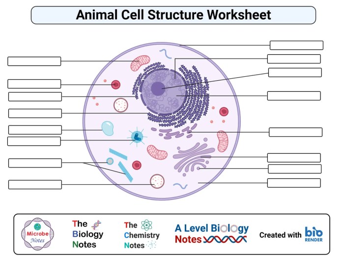
A Coloring Activity Worksheet: The Animal Cell
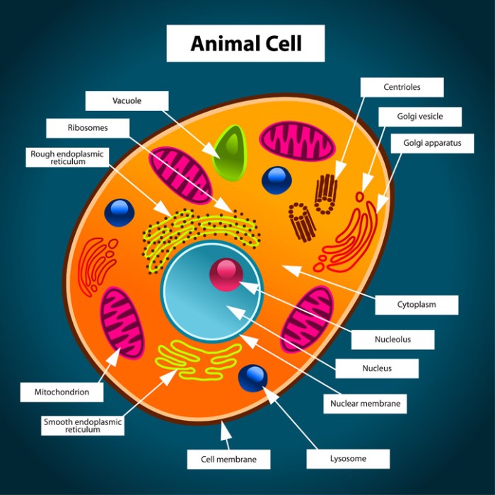
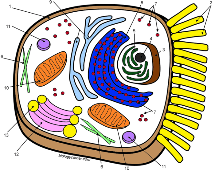
This worksheet focuses on the accurate representation of animal cell organelles and their relative sizes. Students will color each organelle using a specific color, paying attention to its location and approximate size compared to other organelles. Accurate representation is key to understanding the functional relationships between these cellular components.The worksheet depicts a typical animal cell. Students are provided with a labeled diagram outlining the major organelles: the nucleus (large, centrally located, and colored a deep purple to represent its dense genetic material), the rough endoplasmic reticulum (a network of interconnected flattened sacs, colored a light blue to signify its association with ribosomes), the smooth endoplasmic reticulum (a network of tubules, colored a pale green to highlight its distinct function), the Golgi apparatus (a stack of flattened sacs, colored a warm orange to illustrate its processing and packaging role), mitochondria (oval-shaped organelles, colored a vibrant red to represent their energy production), ribosomes (small dots, colored dark gray to represent their role in protein synthesis), lysosomes (small, round organelles, colored a deep magenta to indicate their digestive function), and the cell membrane (the outer boundary, colored a soft yellow to highlight its protective function).
The cytoplasm (the jelly-like substance filling the cell) should be colored a very light yellow-green. The relative sizes of the organelles should be carefully considered and reflected in the coloring, emphasizing the dominance of the nucleus and the comparatively smaller sizes of other organelles. This exercise promotes spatial reasoning and reinforces the understanding of the hierarchical organization within the cell.
Understanding animal cell structures is crucial, and an animal cell coloring answer key provides invaluable assistance in mastering this. However, to appreciate the diversity of life, exploring visuals like those found at ocean animals coloring pages is equally important. This broader perspective reinforces the fundamental principles learned from the answer key, ultimately deepening biological understanding.
The completed coloring should visually represent a lively, functional animal cell, a vibrant testament to the intricate processes occurring within.
Analyzing a Sample Animal Cell Coloring Page

Let’s delve into the fascinating world of animal cell diagrams and explore the common pitfalls students encounter when bringing these microscopic marvels to life with color. Understanding these errors is crucial not only for accurate representation but also for fostering a deeper comprehension of cell structure and function. A seemingly simple coloring activity can reveal surprising misconceptions if not approached carefully.It’s easy to get carried away with the vibrant hues, but accuracy in depicting an animal cell is paramount.
Common mistakes often stem from a lack of understanding of the relative sizes and locations of organelles within the cell. For instance, the nucleus, a prominent feature, is frequently drawn too small or misplaced. Similarly, the mitochondria, often described as the “powerhouses” of the cell, are sometimes overlooked or depicted inaccurately in size and number. The Golgi apparatus, a complex structure involved in protein modification, might be simplified or completely omitted, leading to an incomplete picture.
Another common error is the inconsistent coloring of organelles, leading to a visually confusing and inaccurate representation. Students might also incorrectly color the cytoplasm, a vital component of the cell, or fail to differentiate between the cell membrane and the cytoplasm altogether.
Common Errors in Animal Cell Coloring and Their Corresponding Misconceptions
These inaccuracies in coloring directly translate to misconceptions about the cell’s organization and function. For example, a disproportionately small nucleus might lead a student to underestimate its crucial role in controlling cellular activities and containing genetic material. The omission or misrepresentation of mitochondria could foster a misunderstanding of cellular respiration and energy production. Similarly, ignoring the Golgi apparatus might obscure its vital function in modifying, sorting, and packaging proteins for transport within or outside the cell.
An inaccurate depiction of the cell membrane can lead to misconceptions about its role in regulating the passage of substances into and out of the cell, impacting understanding of processes like osmosis and diffusion. Inconsistent coloring, while seemingly minor, can hinder the ability to visualize the spatial relationships between different organelles and their collective function within the cell.
Correcting Common Mistakes in Animal Cell Coloring
To rectify these common errors, a multi-pronged approach is needed. First, a clear and labeled diagram should be provided as a reference. This diagram should accurately depict the relative sizes and positions of each organelle. For example, the nucleus should be shown as a large, centrally located structure, occupying a significant portion of the cell’s volume. Mitochondria should be illustrated as numerous, elongated organelles scattered throughout the cytoplasm.
The Golgi apparatus should be shown as a series of flattened sacs or cisternae. The cell membrane should be clearly distinguished from the cytoplasm, depicted as a thin, outer boundary. Second, a color-coded key should be included, associating each organelle with a specific color. This ensures consistency and clarity. For instance, the nucleus could be colored purple, the mitochondria blue, the Golgi apparatus green, the cytoplasm a light yellow, and the cell membrane a dark brown.
Third, interactive exercises and activities can help students visualize and understand the 3-D structure of the cell and the spatial arrangement of its organelles. This could involve building a 3-D model of the cell or using virtual reality simulations. Through these combined strategies, students can move from inaccurate representations to a much more accurate and comprehensive understanding of animal cell structure.
Creating a Detailed Animal Cell Diagram with Color-Coding

Embarking on the creation of a detailed animal cell diagram is like assembling a miniature, vibrant city. Each organelle plays a crucial role, and visualizing their individual contributions and spatial relationships is key to understanding the cell’s intricate workings. The use of color-coding elevates this process, transforming a potentially dry exercise into a captivating and memorable learning experience. The vibrant hues help to not only differentiate the organelles but also imprint their identities on our minds.The following section details the creation of such a diagram, emphasizing the importance of clear visual representation and accurate labeling.
We will use color to bring this complex cellular landscape to life, enhancing comprehension and appreciation of this fundamental unit of life.
Animal Cell Organelle Representation and Color Key, Animal cell coloring answer key
Imagine the cell membrane, the bustling city limits, depicted in a rich, deep blue. Within, the nucleus, the city’s command center, shines a brilliant, sunny yellow. The nucleolus, a smaller structure within the nucleus, is a slightly paler, almost lemon yellow. The rough endoplasmic reticulum, a network of roadways dotted with ribosomes (tiny protein factories), is a vibrant, forest green.
The smooth endoplasmic reticulum, responsible for lipid synthesis, is a lighter, spring green. Ribosomes, those industrious protein-makers, are represented by tiny, dark purple dots scattered on the rough ER. The Golgi apparatus, the city’s efficient postal service, is a warm, terracotta orange. Mitochondria, the powerhouses generating energy, are a striking crimson red. Lysosomes, the city’s waste disposal system, are a deep, almost black purple.
The cytoskeleton, a structural framework providing support, is represented by thin, charcoal gray lines extending throughout the cell. Finally, the centrosomes, crucial for cell division, are depicted as small, bright turquoise spheres. This color scheme, carefully chosen for contrast and memorability, aids in visualizing the individual organelles and their interconnections.
Diagram Construction Using HTML Div Tags
To construct our diagram, we utilize the power of HTML <div> tags. Each organelle will receive its own <div>, allowing for precise positioning and clear labeling. Consider this a blueprint for our cellular city, each <div> representing a specific building or infrastructure element. The styling (using CSS, not shown here for brevity) would further refine the visual appeal, ensuring each organelle is clearly distinct and correctly sized relative to the others.
This approach ensures an organized and easily understandable representation. For example, the nucleus, represented by a yellow circle within its <div>, might be positioned centrally. The surrounding organelles, each within their own <div>, are then strategically placed to reflect their spatial relationships within a real animal cell. The use of CSS allows for fine-tuning of size, shape, and position, enabling a highly accurate and visually engaging representation.
FAQ Summary: Animal Cell Coloring Answer Key
What are some common mistakes students make when coloring animal cells?
Common mistakes include inaccurate sizing of organelles, incorrect placement of organelles, and using inappropriate colors that do not reflect the typical appearance of the organelles.
How can coloring activities improve understanding of cell structure?
Coloring helps students visualize the spatial relationships between organelles, reinforces memorization of organelle names and functions, and provides a hands-on learning experience.
Are there specific coloring techniques recommended for accuracy?
Using a legend or color key is crucial. Choosing distinct, easily identifiable colors for each organelle aids in accurate representation and avoids confusion.
How can I use this answer key to assess student understanding?
Compare student work to the provided diagrams and color key, noting accuracy in organelle placement, size, and color. This allows for identification of areas needing further instruction.
