Understanding Animal Cell Structure

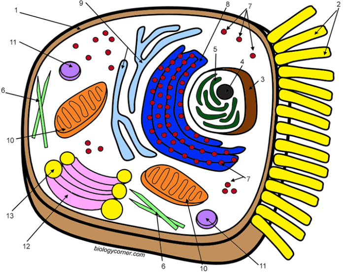
Animal cell coloring answer key – Animal cells are the fundamental building blocks of all animal life, exhibiting a complex and dynamic internal structure. These microscopic units perform a vast array of functions, from energy production and waste removal to protein synthesis and cell division, all orchestrated by specialized internal components called organelles. Understanding the structure and function of these organelles is key to comprehending how animal life thrives.The basic components of a typical animal cell include the cell membrane, cytoplasm, nucleus, and various organelles.
The cell membrane, a selectively permeable barrier, encloses the cell’s contents and regulates the passage of substances in and out. The cytoplasm, a jelly-like substance, fills the cell and houses the organelles. The nucleus, often referred to as the cell’s control center, contains the genetic material (DNA) that dictates the cell’s activities.
Organelle Functions
Each organelle within an animal cell performs a specific role essential for the cell’s survival and function. The mitochondria are responsible for energy production through cellular respiration. Ribosomes synthesize proteins, while the endoplasmic reticulum (ER) modifies and transports these proteins. The Golgi apparatus further processes and packages proteins for secretion or use within the cell. Lysosomes act as the cell’s waste disposal system, breaking down cellular debris and foreign materials.
The cytoskeleton provides structural support and facilitates cell movement.
Specialized Animal Cells and Their Unique Features, Animal cell coloring answer key
While all animal cells share basic components, specialized cells have developed unique features to perform specific tasks.
Understanding animal cell structure through coloring diagrams with an answer key is a great learning tool. For a change of pace, exploring visually appealing sea animals coloring pages can be a fun activity. Then, returning to the animal cell coloring answer key allows for a refreshed and engaged learning experience with cell components.
- Nerve cells (neurons): These cells are characterized by long extensions called axons and dendrites, enabling them to transmit electrical signals throughout the body. This specialized structure facilitates rapid communication between different parts of the organism.
- Muscle cells: Containing a high concentration of contractile proteins like actin and myosin, muscle cells are responsible for movement. The arrangement of these proteins allows for muscle contraction and relaxation, generating force and enabling movement.
- Red blood cells (erythrocytes): These cells are specialized for oxygen transport. Their biconcave disc shape increases surface area for efficient oxygen binding, and the lack of a nucleus maximizes space for hemoglobin, the oxygen-carrying protein. Mature red blood cells in mammals lack a nucleus, which differentiates them from other animal cells.
| Organelle | Function |
|---|---|
| Mitochondria | Energy production |
| Ribosomes | Protein synthesis |
| Endoplasmic Reticulum (ER) | Protein modification and transport |
| Golgi Apparatus | Protein processing and packaging |
| Lysosomes | Waste breakdown |
| Cytoskeleton | Structural support and cell movement |
The cell membrane is a selectively permeable barrier that regulates the passage of substances into and out of the cell.
Coloring as a Learning Tool
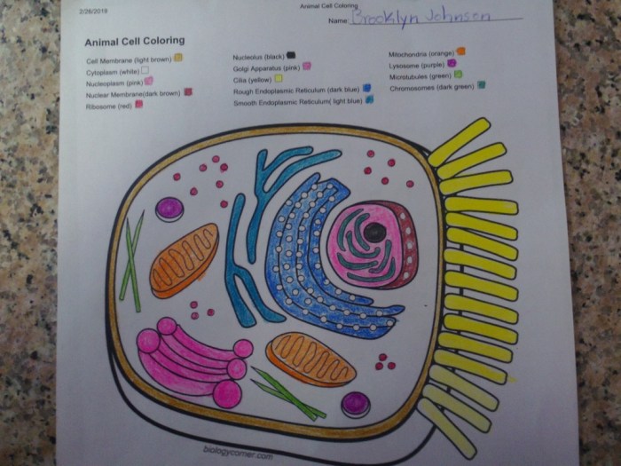
Coloring offers a dynamic approach to understanding complex biological structures like animal cells. It transforms passive learning into an active process, engaging visual and kinesthetic learning styles. This hands-on activity enhances comprehension and retention of information related to cell components and their functions.Coloring facilitates the visualization and memorization of various cell parts. By assigning different colors to organelles like the nucleus, mitochondria, and endoplasmic reticulum, students create visual distinctions.
This color-coding system helps establish a clear mental map of the cell’s organization, making it easier to recall the location and role of each component. The act of coloring reinforces this visual learning, solidifying the connection between the structure and its assigned color.
Color-Coding Techniques for Cell Structures
Color-coding can be strategically implemented to highlight specific relationships within the cell. For instance, using similar shades of blue for the nucleus and ribosomes can emphasize their connection in protein synthesis. Similarly, contrasting colors for the mitochondria and lysosomes can visually distinguish their opposing roles in energy production and waste breakdown, respectively. This thoughtful application of color helps students grasp the interconnectedness of cellular processes.
Integrating Coloring Activities into Science Education
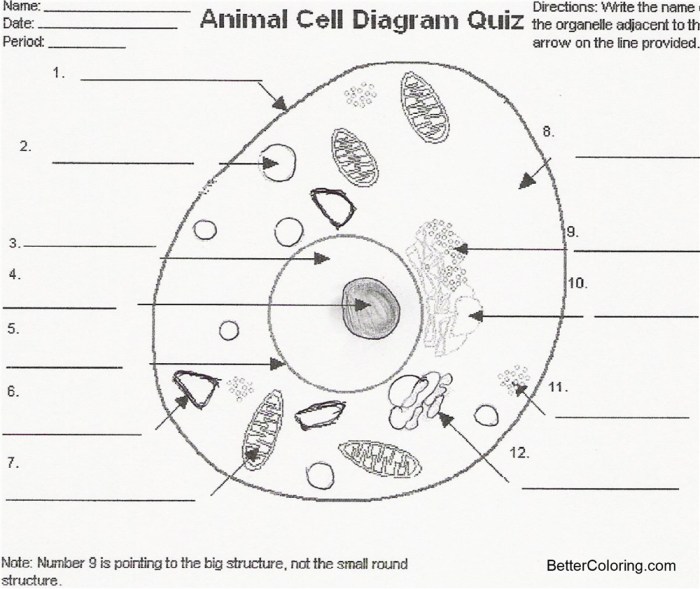
Coloring activities can be seamlessly incorporated into various stages of a lesson. They can serve as an engaging introduction to cell structures, a reinforcing activity after a lecture, or a review tool before an assessment. Providing students with unlabeled diagrams encourages them to actively recall and label the parts themselves before coloring. This reinforces their understanding of both the structure and terminology.
Additionally, group coloring projects can foster collaborative learning, where students discuss and teach each other about different cell components.
| Activity | Description |
|---|---|
| Pre-lesson Warm-up | Students color a basic animal cell diagram, labeling the main organelles. This activates prior knowledge and prepares them for new information. |
| Post-lecture Reinforcement | After learning about specific organelles, students color-code them based on their function (e.g., energy production, protein synthesis). |
| Review and Assessment | Students complete a detailed coloring page, labeling all parts and briefly describing their functions. This serves as a comprehensive review before a test. |
“Coloring engages multiple learning styles, promoting deeper understanding and better retention of information related to cell biology.”
Typical Components of Animal Cell Coloring Pages
Animal cell coloring pages serve as valuable educational tools for visualizing the intricate world within these fundamental units of life. By coloring and labeling key structures, students develop a deeper understanding of the organization and function of animal cells.Coloring pages typically depict a cross-section of a generalized animal cell, showcasing the various organelles responsible for carrying out essential life processes.
These representations, while simplified, effectively convey the spatial relationships between these components.
Organelles Commonly Depicted in Animal Cell Diagrams
Animal cell diagrams often feature a selection of key organelles crucial for cellular function. These include the nucleus, mitochondria, ribosomes, endoplasmic reticulum (both smooth and rough), Golgi apparatus, lysosomes, and the cell membrane.
Visual Representation of Organelles
Organelles are typically represented with distinct shapes and textures to aid identification. The nucleus, often centrally located, is generally depicted as a large, circular or oval structure. Mitochondria, the powerhouses of the cell, are often shown as elongated, bean-shaped structures with internal folds (cristae). Ribosomes, responsible for protein synthesis, are represented as small dots or granules, either free-floating or attached to the endoplasmic reticulum.
The endoplasmic reticulum is portrayed as a network of interconnected membranes, with the rough ER distinguished by the presence of ribosomes on its surface. The Golgi apparatus appears as a stack of flattened, membrane-bound sacs, while lysosomes are typically drawn as small, spherical vesicles. The cell membrane, enclosing the entire cell, is depicted as a thin, continuous line.
Animal Cell Coloring Page Design
The following table presents a simplified animal cell coloring page design with key structural features. Imagine each organelle colored differently for clear distinction. For instance, the nucleus could be purple, the mitochondria orange, the ribosomes blue, the endoplasmic reticulum green, the Golgi apparatus pink, the lysosomes yellow, and the cell membrane a darker blue. The cytoplasm, the jelly-like substance filling the cell, could be a light blue or grey.
| Organelle | Description | Organelle | Description |
|---|---|---|---|
| Nucleus | Contains the cell’s genetic material (DNA) and controls cell activities. Visualized as a large, central, spherical structure. | Mitochondria | Produces energy for the cell through cellular respiration. Depicted as elongated, bean-shaped organelles with internal folds. |
| Ribosomes | Synthesize proteins. Represented as small dots, either free or attached to the endoplasmic reticulum. | Endoplasmic Reticulum (Rough) | Network of membranes studded with ribosomes, involved in protein synthesis and processing. Visualized as interconnected, flattened sacs with attached dots (ribosomes). |
| Endoplasmic Reticulum (Smooth) | Network of membranes involved in lipid synthesis and detoxification. Visualized as interconnected, flattened sacs without ribosomes. | Golgi Apparatus | Processes, sorts, and packages proteins and lipids. Depicted as a stack of flattened, membrane-bound sacs. |
| Lysosomes | Contain enzymes that break down waste materials and cellular debris. Shown as small, spherical vesicles. | Cell Membrane | The outer boundary of the cell, regulating the passage of substances in and out. Represented as a thin, continuous line surrounding the cell. |
Exploring Different Cell Types: Animal Cell Coloring Answer Key

Animal cells exhibit remarkable diversity in structure and function, reflecting their specialized roles within the body. This exploration delves into the unique characteristics of various animal cell types, highlighting their adaptations and contributions to the organism’s overall function.
Structural Comparison of Animal Cells
Different animal cells have evolved unique structural features that enable them to perform their specific tasks effectively. Nerve cells, for instance, possess long, branching extensions called axons and dendrites, facilitating communication over long distances. Muscle cells, responsible for movement, contain contractile proteins arranged in a highly organized manner, allowing for contraction and relaxation. Epithelial cells, forming protective layers and linings, are tightly packed and exhibit various shapes, contributing to their barrier function.
Categorization of Cell Types by Function
Animal cells can be categorized based on their primary functions within the organism. This organization reveals the interconnectedness of different cell types and their contributions to the overall functioning of the body.
| Cell Type | Shape | Key Organelles | Primary Function |
|---|---|---|---|
| Nerve Cell (Neuron) | Elongated with branching extensions (axons and dendrites) | Prominent nucleus, extensive endoplasmic reticulum, numerous mitochondria | Transmission of nerve impulses, communication within the nervous system |
| Muscle Cell (Myocyte) | Elongated, cylindrical, or spindle-shaped | Abundant mitochondria, specialized endoplasmic reticulum (sarcoplasmic reticulum), myofibrils (contractile proteins) | Contraction and relaxation, enabling movement |
| Epithelial Cell | Various shapes (cuboidal, columnar, squamous) | Tight junctions between cells, prominent cytoskeleton | Protection, lining of surfaces, secretion, absorption |
| Red Blood Cell (Erythrocyte) | Biconcave disc | Lacks nucleus and most organelles in mature form, contains hemoglobin | Oxygen transport throughout the body |
Visualizing Cellular Processes

Coloring diagrams provides a dynamic and engaging way to understand complex cellular processes. By actively coloring specific components involved in these processes, students can visualize the steps and interactions, leading to a deeper understanding of how these intricate systems function. This hands-on approach reinforces learning and helps solidify the connection between structure and function within the cell.
Protein Synthesis Coloring Activity
This activity focuses on visualizing the process of protein synthesis, from DNA transcription to translation and protein assembly. The coloring page would depict the key organelles and molecules involved, including the nucleus, ribosomes, mRNA, tRNA, and amino acids. Different colors will be assigned to each component to highlight their distinct roles.
Step 1: Transcription. Color the DNA (deoxyribonucleic acid) double helix within the nucleus blue. Then, color the mRNA (messenger ribonucleic acid) molecule, which is being transcribed from the DNA, red. This step represents the creation of an mRNA copy of a specific gene.
Step 2: mRNA Transport. Color the mRNA molecule as it moves out of the nucleus and into the cytoplasm yellow. This visualizes the transport of the genetic information from the nucleus to the site of protein synthesis.
Step 3: Translation at the Ribosome. Color the ribosome green. Ribosomes are the protein synthesis machinery of the cell. Color the tRNA (transfer RNA) molecules, which carry specific amino acids, purple. Show these tRNA molecules interacting with the mRNA at the ribosome.
Step 4: Amino Acid Chain Formation. Color the growing chain of amino acids orange. This chain represents the polypeptide that will eventually fold into a functional protein. Each amino acid is brought to the ribosome by a tRNA molecule and added to the growing polypeptide chain based on the mRNA sequence.
Step 5: Protein Folding. Color the final folded protein brown. This represents the completed protein, now in its functional three-dimensional structure, ready to perform its specific role within the cell.
Creating Educational Resources
Animal cell coloring pages offer a dynamic approach to learning about cell biology. Beyond simply filling in colors, these pages can serve as a foundation for interactive lessons that promote deeper understanding of cellular structures and functions. By incorporating discussions, research activities, and creative projects, educators can transform coloring pages into powerful educational tools.Interactive learning materials can be developed using animal cell coloring pages as a starting point.
This approach encourages active participation and reinforces learning through visual and kinesthetic activities.
Interactive Learning Activities with Animal Cell Coloring Pages
Coloring pages can be integrated into a broader lesson plan to enhance understanding of animal cell biology. This integration allows students to connect visual representations with conceptual knowledge.
- Begin by having students color the different organelles within an animal cell, using a provided key or research materials to identify and label each component.
- Facilitate a discussion about the function of each organelle, encouraging students to explain their roles in the cell’s overall operation. This can be achieved through group work, presentations, or question-and-answer sessions.
- Extend the learning experience by having students conduct further research on specific organelles or cellular processes. They can create presentations, write reports, or build models to demonstrate their understanding.
Designing a Lesson Plan
A well-structured lesson plan can effectively incorporate coloring activities, discussions, and research to maximize learning outcomes. This approach provides a framework for a comprehensive learning experience.
| Activity | Description | Materials |
|---|---|---|
| Introduction (10 minutes) | Brief overview of animal cells and their importance. | Slideshow, diagrams |
| Coloring Activity (20 minutes) | Students color and label the organelles of an animal cell. | Animal cell coloring pages, colored pencils, labels |
| Group Discussion (15 minutes) | Students discuss the function of each organelle and its role in the cell. | Whiteboard, markers |
| Research and Presentation (30 minutes/homework) | Students research a specific organelle or cellular process and prepare a presentation. | Internet access, presentation software |
| Wrap-up (5 minutes) | Review key concepts and address any remaining questions. | None |
Creating 3D Models
Building three-dimensional models provides a tangible representation of animal cells, enhancing spatial understanding of cellular structures.Students can use various materials like clay, foam balls, pipe cleaners, and construction paper to create a 3D model of an animal cell. Each organelle can be represented using a different material and color, allowing students to visualize the arrangement and relative sizes of these components within the cell.
For instance, the nucleus could be represented by a larger foam ball, while smaller beads could represent ribosomes. Labels can be added to identify each organelle and its function.
A 3D model allows students to interact with the cell structure in a more tangible way, reinforcing their understanding of the spatial relationships between organelles.
